Hokkaido University researchers have figured out how to forestall gold nanoparticles from clustering, which could help towards their utilization as an enemy of malignant growth treatment.
Connecting ring-molded engineered mixes to gold nanoparticles causes them hold their fundamental light-retaining properties, Hokkaido University specialists report in the diary Nature Communications.
Metal nanoparticles have novel light-engrossing properties, making them intriguing for a wide scope of optical, electronic and biomedical applications. For instance, whenever conveyed to a tumor, they could respond with applied light to murder malignant tissue. An issue with this methodology, however, is that they effectively bunch together in arrangement, losing their capacity to retain light. This amassing occurs in light of an assortment of components, including temperature, salt fixation and sharpness.
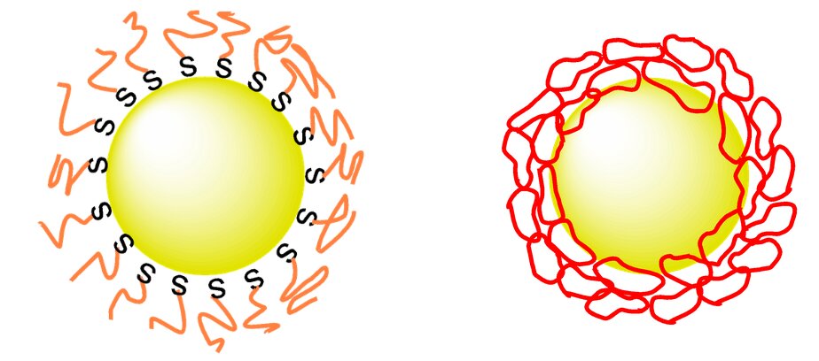
Researchers have been attempting to discover approaches to guarantee nanoparticles stay scattered in their objective surroundings. Covering them with polyethylene glycol, also called PEG, has been moderately fruitful at this on account of gold nanoparticles. Stake is biocompatible and can keep gold surfaces from bunching together in the research facility and in living organic entities, however upgrades are as yet required.

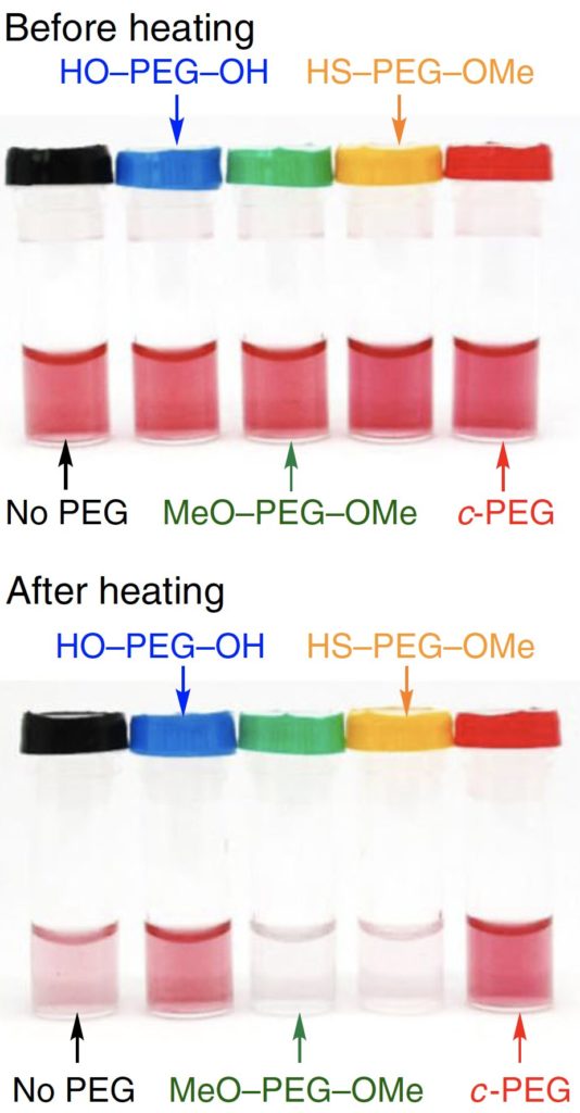
Gold nanoparticles suspended in various PEG arrangements. In the wake of warming, gold nanoparticles in c-PEG had the most elevated scattering security, which corresponds to the shading power. Credit: Yubo Wang et al, Nature Communications, November 30, 2020
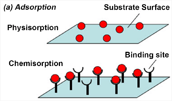
Applied scientific expert Takuya Yamamoto and associates at Hokkaido University, The University of Tokyo, and Tokyo Institute of Technology found that blending gold nanoparticles with ring-formed PEG, as opposed to the typically straight PEG, altogether improved scattering. The ‘cyclic-PEG’ (c-PEG) appends to the surfaces of the nanoparticles without framing synthetic bonds with them, a cycle called physisorption. The covered nanoparticles stayed scattered when frozen, freeze-dried and warmed.

The group tried the c-PEG-shrouded gold nanoparticles in mice and found that they cleared gradually from the blood and gathered better in tumors contrasted with gold nanoparticles covered with direct PEG. In any case, amassing was lower than wanted levels, so the specialists prescribe further examinations to adjust the nanoparticles for this reason.
Partner Professor Takuya Yamamoto is important for the Laboratory of Chemistry of Molecular Assemblies at Hokkaido University, where he considers the properties and utilizations of different cyclic synthetic mixes.

Dipanjan Pan, educator of substance, biochemical, and ecological designing at UMBC, and associates distributed a fundamental report in Nature Communications that shows unexpectedly a strategy for biosynthesizing plasmonic gold nanoparticles inside disease cells, without the requirement for regular seat top lab techniques. It can possibly outstandingly extend biomedical applications.
Traditional research facility based blend of gold nanoparticles require ionic antecedents and decreasing specialists exposed to fluctuating response conditions, for example, temperature, pH, and time. This prompts variety in nanoparticle size, morphology and functionalities that are straightforwardly related to their disguise in cells, their home time in vivo, and freedom. To keep away from these vulnerabilities, this work shows that biosynthesis of gold nanoparticles can be accomplished proficiently straightforwardly in human cells without requiring ordinary research center strategies.
The analysts inspected how different disease cells reacted to the acquaintance of chloroauric corrosive with their cell microenvironment by shaping gold nanoparticles. These nanoparticles created inside the cell can possibly be utilized for different biomedical applications, remembering for X-beam imaging and in treatment by annihilating unusual tissue or cells.

In the paper, Pan and his group portray their new strategy for creating these plasmonic gold nanoparticles inside cells in minutes, inside a cell’s core, utilizing polyethylene glycol as a conveyance vector for ionic gold. “We have built up a one of a kind framework where gold nanoparticles are decreased by cell biomolecules and those can hold their usefulness, including the ability to control the leftover group to the core,” says Pan.
They additionally attempted to additionally show the biomedical capability of this methodology by actuating in-situ biosynthesis of gold nanoparticles inside a mouse tumor, trailed by photothermal remediation of the tumor. Dish clarifies that the mouse study exemplified how “the intracellular arrangement and atomic relocation of these gold nanoparticles presents an exceptionally encouraging methodology for drug conveyance application.”
“Gold is the quintessential respectable component that has been utilized in biomedical applications since its first colloidal combination over three centuries back,” Pan notes. “To value its potential for clinical application, nonetheless, the most testing research in front of us will be to discover new strategies for creating these particles with positive reproducibility with functionalities that can advance productive cell authoritative, leeway, and biocompatibility and to survey their drawn out term consequences for human wellbeing. This new investigation is a little yet significant advance toward that all-encompassing objective.”
